Background: CNS invasion in aggressive lymphoma (including diffuse large B-cell [DLBCL], double-hit [DHL], Burkitt [BL] and plasmablastic lymphoma [PBL]) remains a major diagnostic and therapeutic challenge. Parenchymal CNS invasion requires a brain biopsy to diagnose, and conventional CSF evaluations lack sensitivity. Furthermore, determining which patients (pts) without CNS disease at diagnosis might be at high risk of future CNS relapse, and thus would most benefit from aggressive CNS prophylaxis, is difficult. Predictors that use clinical variables (e.g. the CNS-IPI) can select a high-risk group with ~10% risk of CNS relapse, but ~50% of events occur in low/intermediate-risk groups. We evaluated a novel CSF assay that detects clonotype-specific cell-free DNA (cfDNA) by next-generation sequencing (NGS) combined with multiplex PCR (Chang T et al, BMC Cancer 2020). This assay can identify minimal residual disease (MRD) in the plasma of pts with DLCBL and can predict systemic relapse (Roschewski et al, Lancet Oncol 2015).
Methods: To examine the diagnostic sensitivity of the NGS-MRD assay in the CSF, we prospectively collected CSF from 6 pts with lymphomas who had known parenchymal or leptomeningeal CNS invasion. We also tested stored DNA from historical CSF samples of 8 pts with CNS lymphoma who tested negative by conventional IGH-PCR. We then enrolled 19 pts with newly diagnosed high-risk lymphomas without CNS invasion to examine the prognostic value of the NGS-MRD assay in the CSF collected at diagnosis. In the two-step assay, tumor-specific clonotype sequences were first identified from genomic DNA in primary tumor specimens using NGS of rearranged IGK, IGH (VJ or DJ), and IGL loci. Tumor-specific clonotypes were then selected for tracking in the CSF (split into acellular CSF fluid and cell pellet) and in paired plasma samples. The assay quantitated copy numbers of each tracked sequence per mL of acellular CSF or per 106 cells in the pellet, as well as tumor clonotype frequency (%) per all B-cells.
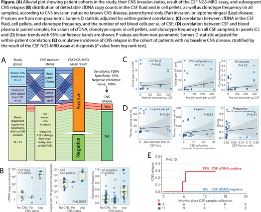
Results: The NGS-MRD detected tumor-specific clonotype in 100% of CSF samples from pts with known CNS lymphoma (n=6; Fig A), including pts with isolated parenchymal-only CNS disease that was not detected by CSF cytology, flow cytometry, or IGH-PCR. For pts with CNS parenchymal disease, median cfDNA copy count in the CSF fluid was 2 /mL (range, 0.4-929) and median clonotype frequency was 9.0% (range, 0.03-68.9%). For pts with leptomeningeal disease, these values were 2233 /mL (1.2-5620) and 37% (1.7-98.4%), respectively (Fig. B). Furthermore, the NGS-MRD assay detected tumor clonotypes in DNA isolates from 6 historical CSF samples (collected in 2014-2018) of pts with known CNS disease, in which lymphoma was not detectable by conventional IGH-PCR (2 additional historical samples failed DNA quality check).
Among 19 newly diagnosed pts with no known CNS invasion (median age 57, 42% women, median LDH 352 IU/L), 12 (63%) had DLBCL with high risk of CNS relapse by virtue of high CNS-IPI or epidural tumor, and 7 had other high-risk histologies (2 DHL, 2 BL, HIV+ PBL, and 2 lymphoblastic leukemias). The CSF NGS-MRD assay was positive for tumor-specific clonotype in 8 pts (42%). Median cfDNA copy count in the CSF fluid was 3 /mL (range, 0.9-15.6) and median clonotype frequency was 18.6% (range, 1.4-28.5%). We observed no significant correlation between the amount of cfDNA and number of red cells /mL of CSF (a measure of potential CSF contamination by blood during collection procedure; Fig. C) or between the amount of cfDNA in plasma and in CSF (Fig. D). With median follow-up of 11 months, no pts with negative baseline NGS-MRD assay in CSF relapsed, whereas 2 of 10 with a positive assay had a CNS relapse (12-month incidence, 29%; Fig. E), despite both pts having negative CNS imaging, CSF cytology, flow cytometry, and IGH-PCR at diagnosis.
Conclusions: In this proof-of-concept study, the NGS-MRD CSF assay showed 100% sensitivity for diagnosing intraparenchymal CNS invasion in aggressive lymphomas that were not detected in the CSF using conventional methods. In a prospective cohort of newly diagnosed pts without baseline CNS invasion but at high clinical risk for CNS relapse, the assay identified lymphoma-derived cfDNA in 42% of cases. The 100% sensitivity and 100% negative predictive value of the assay for subsequent CNS relapse warrants further prospective evaluation as a potential tool for personalizing CNS-directed prophylaxis.
Olszewski:Spectrum Pharmaceuticals: Research Funding; TG Therapeutics: Research Funding; Adaptive Biotechnologies: Research Funding; Genentech, Inc.: Research Funding. Mullins:Adaptive Biotechnologies: Current Employment, Other: shareholder.
Author notes
Asterisk with author names denotes non-ASH members.

This icon denotes a clinically relevant abstract


This feature is available to Subscribers Only
Sign In or Create an Account Close Modal